How Does Breast Cancer Look Like In Ultrasound - Here's what to do if you think you've found a lump in your ... : If your breast tissue is too dense for a mammogram.
Dapatkan link
Facebook
X
Pinterest
Email
Aplikasi Lainnya
How Does Breast Cancer Look Like In Ultrasound - Here's what to do if you think you've found a lump in your ... : If your breast tissue is too dense for a mammogram.. This appears most commonly as streaking, known as linear enhancement. What does a solid mass look like in an ultrasound breast image? To look more closely at a. My doctor found a lump in my breast during my yearly mammogram and then brought in an ultrasound technician to follow up with an ultrasound for a closer look. The doctor must be sure it's a cyst to know it's not cancer.
In the table the typical ultrasound findings are listed (click to enlarge). Other ultrasound findings that suggest breast cancer include: This is because it may miss some early signs of cancer. A breast ultrasound is most often done to find out if a problem found by a mammogram or physical exam of the breast may be a cyst filled with fluid or a solid tumor. Solid lesions can be a little brighter or darker than the surrounding tissue, and the way to evaluate these on ultrasound is to look closely at the margins or the outer edges of the nodule.
What is a fibroadenoma? Should I have it removed? - My ... from mybreastmyhealth.com Ultrasound is frequently used to evaluate breast abnormalities that are found with screening mammography or diagnostic mammography or during a physician performed clinical breast exam.ultrasound allows significant freedom in obtaining images of the. You may notice dimpling or pitting, and the skin on your breast. There is a slight increase in the density in the right breast compared with the left. We will discuss each of these findings in more detail in a moment. What does a solid mass look like in an ultrasound breast image? This is primarily a benign mammary dysplasia and papilloma ducts. They can also look the same on a mammogram. Breast cancer usually makes or presents as a mass or tumor or a lump.
You can get dressed straight after the ultrasound.
A breast ultrasound is a scan that uses penetrating sound waves that do not affect or damage the tissue and cannot be heard by humans. Tumors are likely to be smaller when doctors detect them early, which can. Moose & doc breast cancer, 21 may 2018. 1 send thanks to the doctor They can also look the same on a mammogram. They cause a large amount of the sound waves projected at them to bounce back towards the machine. While it may look like a fuzzy, spotty television screen with different shades of grey to a patient, the ultrasound technician and the radiologist use these images to diagnose masses and tumors. Physical examination and mammogram can be more accurate in some settings. Dcis on mri may create an area of irregular enhancement of the mri dye into the breast. Tumor size is an important factor in breast cancer staging, and it can affect a person's treatment options and outlook. Ultrasound is only one means of evaluation of the breast. Other ultrasound findings that suggest breast cancer include: A cyst and a solid mass can feel the same.
With ultrasound, the radiologist will probably be trying to get a sense of the internal texture of the breast lesion and surrounding area. While it may look like a fuzzy, spotty television screen with different shades of grey to a patient, the ultrasound technician and the radiologist use these images to diagnose masses and tumors. You can get dressed straight after the ultrasound. Dcis on mri may create an area of irregular enhancement of the mri dye into the breast. A rash isn't the only visual symptom of inflammatory breast cancer.
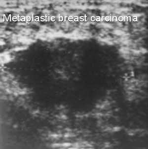
Metaplastic breast cancer - Moose and Doc from breast-cancer.ca Ultrasound characterization of breast masses. indian journal of radiology and imaging. Not all suspicious breast lesions will direct in appearance and ultrasound diagnosis. Cysts, tumors, and growths will appear as dark areas on the scan. Dcis on mri may create an area of irregular enhancement of the mri dye into the breast. It is usually preceded by an injury to the breast from a car accident or sports injury. This is primarily a benign mammary dysplasia and papilloma ducts. This appears most commonly as streaking, known as linear enhancement. The breast tissue deflects these waves causing echoes, which a computer uses to paint a picture of what's going on inside the breast tissue (no radiation is involved).
Ultrasound is frequently used to evaluate breast abnormalities that are found with screening mammography or diagnostic mammography or during a physician performed clinical breast exam.ultrasound allows significant freedom in obtaining images of the.
The images that a breast ultrasound produces are in black and white. Solid lesions can be a little brighter or darker than the surrounding tissue, and the way to evaluate these on ultrasound is to look closely at the margins or the outer edges of the nodule. Ultrasound characterization of breast masses. indian journal of radiology and imaging. A breast ultrasound is most often done to find out if a problem found by a mammogram or physical exam of the breast may be a cyst filled with fluid or a solid tumor. By far the most common abnormalities in the breast, which usually present as a lump in the breast are cysts, fibroadenomas, breast cancer and palpable glandular tissue. This appears most commonly as streaking, known as linear enhancement. They can also look the same on a mammogram. Breast cancer is among the most common causes of cancer deaths today, coming fifth after lung, stomach, liver and colon cancers. Ultrasound is very accurate in such cases and may be used to do a biopsy. In most cases, breast cancers develops inside the presence of precancerous changes. This is primarily a benign mammary dysplasia and papilloma ducts. On ultrasound, a breast cancer tumor is often seen as hypoechoic, has irregular borders, and may appear spiculated. The dye collection in the breast can also look clumpy or appear in a section of the breast, depending on the involvement of dcis.
This is primarily a benign mammary dysplasia and papilloma ducts. The doctor must be sure it's a cyst to know it's not cancer. There is a slight increase in the density in the right breast compared with the left. Breast cancer is among the most common causes of cancer deaths today, coming fifth after lung, stomach, liver and colon cancers. A specialist looks at the ultrasound pictures.
Detecting breast cancer: Sonograms, 3-D mammography get ... from www.trbimg.com Mammography is the least sensitive imaging. By far the most common abnormalities in the breast, which usually present as a lump in the breast are cysts, fibroadenomas, breast cancer and palpable glandular tissue. The images that a breast ultrasound produces are in black and white. Ultrasound imaging allows better evaluation of the status of the axillary lymph nodes in patients with ibc, an important step in determining extent of disease prior to initiation of chemotherapy. This breast cancer ultrasound image shows changes related to breast cancer that are not seen as microcalcifications or a mass or lump. 1 send thanks to the doctor Below are images of dcis on breast ultrasound. A breast ultrasound is a scan that uses penetrating sound waves that do not affect or damage the tissue and cannot be heard by humans.
Moose & doc breast cancer, 21 may 2018.
Among all cancers in women, he takes the initial place. If a solid lump shows on the scan you might need to have. The dye collection in the breast can also look clumpy or appear in a section of the breast, depending on the involvement of dcis. In most cases, breast cancers develops inside the presence of precancerous changes. We will discuss each of these findings in more detail in a moment. What does the doctor look for on a mammogram? cancer.org. In the table the typical ultrasound findings are listed (click to enlarge). My doctor found a lump in my breast during my yearly mammogram and then brought in an ultrasound technician to follow up with an ultrasound for a closer look. This appears most commonly as streaking, known as linear enhancement. On ultrasound, a breast cancer tumor is often seen as hypoechoic, has irregular borders, and may appear spiculated. If you're younger than 25. A specialist looks at the ultrasound pictures. Breast ultrasound is not usually done to screen for breast cancer.
Keeping things clean and simple helps to inspire trust—something you need on your side in this occasionally volatile industry. Cryptocurrency might be complex, but that doesn't mean your logo layout should be. A simple layout makes your logo more scalable, but you may also choose to create a few variations to make sure your crypto logo looks its best on every trading … This free logo maker tool is a good choice for a variety of reasons, including: Your crypto logo should represent your brand, help people remember you and provide insight into your services. Aclk Sa L Ai Dchcsewiro4lg Mr0ahxdp9ukhe1tbmkyababggj3cw Sig Aod64 3lttkjc6w H 1pdu P0443xt0pyq Adurl Ctype 5 from You'll be presented with 100s of custom logo mockups based on your preferences. Cryptocurrency in general has become very popular as a means of conducting business t...
Sears Card Make Payment - Review: Sears MasterCard - The Shocking 25.24% APR ... - Be sure to bring the credit card bill, as well as cash or a check for the amount you wish to pay. . Just search on google, sears credit card near you. 1 space only between each word or character. Pay sears credit card bill; If you are a cardholder and the payment due date is near and you are wondering how you will pay the bill, read our guide and you will get complete information. Pay online here pay my sears credit card on next landing page select your card option you will be prompted to login, use your existing account online credentials as user id and password hit sign on and follow the prompts. We accept the following gift cards: Find one sears store near you and then go there then make the payment. If you're looking for a new credit card, check out the great member benefits available with a sears card or sears mastercard® card. La próxima vez que inicies sesión, el contenido d...
20.09.2013 · cryptocurrency news (ccn) offers breaking news, analysis, price charts & more on the most popular cryptocurrencies such as bitcoin, litecoin, ethereum & ripple & emerging cryptocurrencies such as monero, stellar, dash & eos. Cryptocurrencies such as bitcoin and ethereum enjoy high levels of liquidity and trade at similar rates regardless of which specific cryptocurrency exchange you're looking at. In a recent note, the company's chief executive highlighted why eth may reach $8,000 by the end of the year. Ethereum is the second cryptocurrency ever created, except it works on a completely algorithm compared to bitcoin. Full list, more then 1500 cryptos can be found, by clicking "load more" button at the bottom of the chart, or just type any cryptocurrency symbol or name in the search box at the top of the chart. Cryptocurrency concept -...


Komentar
Posting Komentar